Pages

Thursday, September 22, 2011

Child Sexual Abuse

2 comments
 
http://img.medscape.com/pi/emed/ckb/pediatrics_development/912185-912186-915841-915943tn.jpg
Child Abuse and Neglect, Sexual Abuse 

When most people think of Child,Neglect,Sexual,Abuse, what comes to mind is usually basic information that's not particularly interesting or beneficial. But there's a lot more to Child,Neglect,Sexual,Abuse than just the basics.

Once you begin to move beyond basic background information, you begin to realize that there's more to Child,Neglect,Sexual,Abuse than you may have first thought. 

Background
Child sexual abuse (CSA) refers to the use of children in sexual activities when, because of their immaturity and developmental level, they cannot understand or give informed consent. A wide range of activities is included in sexual abuse, including contact and noncontact activities. Contact activities included are sexualized kissing, fondling, masturbation, and digital and/or object penetration of the vagina and/or anus, as well as oral-genital, genital-genital, and anal-genital contact. Noncontact activities include exhibitionism, inappropriate observation of child (eg, while the child is dressing, using the toilet, bathing), the production or viewing of pornography, or involvement of children in prostitution.
The sexual activities are imposed on the child and represent an abuse of the caregiver's power over the child.
Genital examination of girl in frog-leg supine position after genital trauma. Examination reveals suture in place at 6-o'clock position to stop bleeding from injury. Hymenal edge is irregular and asymmetric. Photo courtesy of Carol D. Berkowitz, MD.
Since the mid 1970s, health care professionals have paid serious attention to sexual abuse of children. Despite the recognition of the clinical importance of sexual abuse of children, some pediatricians may not feel adequately prepared to perform medical evaluations. However, pediatricians are often in trusted relationships with patients and families and are in an ideal position to offer essential support to the child and family. Several paradigms have been proposed to help professionals understand the events that surround the sexual maltreatment of children.
Preconditions for sexual abuse
Motivation of perpetrator: The perpetrator is willing to act on impulses associated with sexual arousal related to children.
Overcoming internal inhibitions: The perpetrator ignores internal barriers against sexually abusing children.
Overcoming external inhibitions: The perpetrator is able to bypass the typical barriers in the caregiving environment that normally serve to impede the sexual misuse of children.
•    Overcoming child resistance: The perpetrator is able to manipulate the child to the point of involving the child in the sexual activity.
Longitudinal progression of sexual abuse
Engagement: The perpetrator begins relating to the child during nonsexual activities to gain the child's trust and confidence.
Sexual interaction: The perpetrator introduces sexual activities into the relationship with the child; the perpetrator often begins with noncontact types of activities and, over time, progresses to more invasive forms of contact activities.
Secrecy: The perpetrator attempts to maintain access to the child and to avoid disclosure of the abuse by coercing the child to keep the activities hidden. Coercion to keep the secret can be explicit (eg, threatening the child or the child's family's safety) or it can be implicit (eg, manipulation of the child's trust to create a fear of losing the "friendship" or "attention" should the truth become known to others).
Disclosure: Sexual abuse can become known to others either accidentally, when a symptom from the maltreatment or a third party witnessing the abuse leads to an evaluation, or can be purposeful, as when the child reveals the abuse that is taking place and seeks help.
Suppression: The tumult that occurs after the disclosure prompts the people in the child's caregiving environment to think that they are unable to support the child; thus, these people exert pressure on the child to recant what the child has told in order to go back to the perceived "stable" situation that existed prior to the disclosure.
Sexual abuse typically presents as a pattern of maltreatment that occurs over time.
Traumagenic dynamics model
Traumatic sexualization: The child's sexual feelings and attitudes are shaped in a developmentally inappropriate and interpersonally dysfunctional manner. The child learns that sexual behavior may lead to rewards, attention, or privileges. Traumatic sexualization may also occur when the child's sexual anatomy is given distorted importance and meaning.
The child manifests symptoms of fear, anxiety, and impaired coping.
•    Stigmatization: The child's self-image incorporates negative connotations and is associated with words such as bad, awful, shameful, and guilty.
Pathophysiology
The evaluation for suspected sexual abuse may be complicated and is often not straightforward. Frequently, nonspecific behavioral changes are the presenting symptoms prompting an evaluation and leading the health care provider to consider sexual abuse as a possible diagnosis. These nonspecific behaviors are not diagnostic of sexual maltreatment and may be observed in other situations where the child manifests stress as well. Nonspecific behavior changes that warrant consideration of the possibility of sexual abuse may include (1) sexualized behaviors, (2) phobias, (3) sleep disturbances, (4) changes in appetite, (5) change in or poor school performance, (6) regression to an earlier developmental level, (7) running away, (8) truancy, (9) aggressiveness and acting out behaviors, and/or (10) social withdrawal, sadness, or symptoms of depression.
For the purposes of this discussion, the differential diagnosis for each of the following 4 genital findings known to be associated with child sexual abuse is discussed.
Genital bleeding

http://img.medscape.com/pi/emed/ckb/pediatrics_development/912185-912186-915841-915937tn.jpg
Blood-tinged vaginal or urethral discharge initially may be confused with frank bleeding. Differential diagnoses are as follows:
http://img.medscape.com/pi/emed/ckb/pediatrics_development/912185-912186-915841-915938tn.jpg
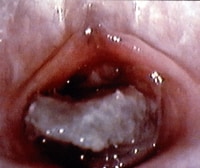
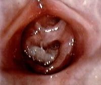
Local factors, such as injury (either accidental or nonaccidental) and/or foreign body irritation (eg, small toy parts, clumped toilet tissue [see the images below])  Prepubertal girl with foul-smelling bloody discharge. The foreign body is lodged in vagina and appears to be toilet tissue that is colonized with bacteria, causing a vulvovaginitis. The foreign body was dislodged with gentle water flushing during examination. Photo courtesy of Carol D. Berkowitz, MD. Genital examination of prepubertal girl with foul-smelling bloody discharge. The foreign body was dislodged with gentle water flushing during examination. Infections, including sexually transmitted diseases (STDs), fungal infections, and/or nonspecific vulvovaginitis
Structural abnormalities, such as vulvar hemangioma
Vaginal discharge
Differential diagnoses are as follows:
Local irritation from abusive sexual contact, foreign body, chemical irritants, and restrictive clothing
Infections, including STDs, fungal infections, nonspecific vulvovaginitis, group A streptococci, Staphylococcus aureus, Haemophilus influenzae, and Mycoplasma species
Physiologic leukorrhea
Anogenital bruising
Differential diagnoses are as follows:
Local injury, including straddle injury, accidental impaling injury, accidental blunt trauma, and abusive injury
Dermatologic conditions, such as mongolian spots, lichen sclerosis, and vascular nevi
Anogenital redness
Local irritation from sexual abuse, poor hygiene, restrictive clothing, and chemicals
Anatomic/structural factors such as perianal fissuring and rectal prolapse
Dermatologic conditions, such as lichen sclerosus, psoriasis, and dermatitis (atopic, contact, seborrhea)
Systemic manifestations of other disorders, such as Crohn disease, Kawasaki syndrome, and Stevens-Johnson syndrome
United States
Prevalence
Professionals conservatively use child sexual abuse prevalence estimates of 20% in women and 5-10% in men. A classic prevalence study of New England male and female college students done by Finkelhor (1984), which used a definition that included both contact and noncontact abuse with older perpetrators and children younger than 17 years, revealed that 19.2% of female students (1 in 5 women) and 9% of male students (1 in 10 men) reported sexual misuse during their childhoods.[4]
Incidence
According to the US Federal Government's official report, Child Maltreatment 2006, approximately 905,000 children were determined to be victims of child abuse; the overall child maltreatment rate was 12.1 cases per 1,000 children.[5] Overall, in 2006, the 905,000 substantiated cases emerged from approximately 3.3 million reports of alleged child abuse and neglect, involving about 6 million children. In addition to the 8.8% of substantiated cases of sexual abuse, an additional 16% were substantiated for physical abuse, and 64.1% were substantiated for child neglect.
In 2009, release of the Fourth National Incidence Study of Child Abuse and Neglect (NIS-4) is expected and eagerly awaited.[6] Prior to the NIS-4's release, older data remain available from the previously congressionally mandated Third National Incidence Study of Child Abuse and Neglect (NIS-3). In 1993, this study reported an estimated sexual abuse incidence rate of 3.2 cases per 1000 children (or a total of 217,000);[7] this represented 29% of the total number of children known to have been abused. NIS-3 used a definition that subsumed a range of behaviors, including intrusion, genital molestation, exposure, inappropriate fondling, and unspecified sexual molestation.
At present, the NIS-3 is the single most comprehensive source of information about the current incidence of child abuse and neglect in the United States and is based on a nationally representative sample. With sexual abuse, the number of undisclosed incidents is believed to be large due to the stigma and criminal behavior involved.
The 1993 NIS-3 incidence figure of 3.2 cases per 1000 children represents a statistically significant (68%) increase from the 1986 Second National Incidence Study of Child Abuse and Neglect (NIS-2) incidence of 1.9 per 1000 children. In part, this difference is due to increased recognition of sexual abuse in the pediatric population. Finkelhor and Jones (2008) at the Crimes Against Children Research Center have been tracking the trends in child maltreatment statistics collected by the Federal Government and have found a national decline in the incidence of both physical and sexual abuse that began in the middle the 1990s and continues through the early 2000s.[8]
However, no decline was found in the rate of child neglect. Specifically, child sexual abuse substantiations have seen a 53% downward trend from the peak annual incidence observed in 1992 (see image below). From 2005-2006, substantiated child sexual abuse cases declined 5%.
http://img.medscape.com/pi/emed/ckb/pediatrics_development/912185-912186-915841-1597776tn.jpg
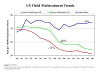
US child maltreatment trends.
Finkelhor and Jones have explored the potential reasons for the decline in child sexual abuse cases and have focused on factors that may be impacting the actual incidence as well as factors that may be influencing the reporting and investigation of reported cases, which may then downstream impact the number of substantiated cases (see image below).
http://img.medscape.com/pi/emed/ckb/pediatrics_development/912185-912186-915841-1597784tn.jpg
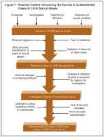
Possible factors influencing the decline in substantiated cases of child sexual abuse.
Optimistically, prevention efforts, incarceration, and treatment of perpetrators (along with other societal factors) may actually be decreasing the number of children who are harmed by sexual abuse. Numerous psychological and medical consequences have been described as associated with sexual abuse. Psychological disorders are reported as having an increased incidence in those who have been abused sexually and include depression, eating disorders, anxiety disorders, substance abuse, somatization, posttraumatic stress disorder (PTSD), dissociative disorders, psychosexual dysfunction in adulthood, and numerous interpersonal problems, including difficulties with issues of control, anger, shame, trust, dependency, and vulnerability.
PTSD and its relationship to sexual abuse have received considerable professional attention. Note that no universal short-term or long-term impact of sexual abuse has been identified, and the presence or absence of various symptoms or conditions does not indicate nor disprove the occurrence of sexual abuse.
Medical sequelae of sexual abuse include numerous medical conditions, including functional GI disorders (eg, irritable bowel syndrome, dyspepsia, chronic abdominal pain), gynecologic disorders (eg, chronic pelvic pain, genital or anal tears), and various forms of somatization involving neurologic conditions and pain syndromes. Additionally, children may contract STDs via sexual abuse, and postpubertal females may become pregnant.
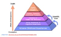
In groundbreaking work, Felitti et al have explored the connection of exposure to childhood abuse and household dysfunction to subsequent health risks and the development of illness in adulthood in a series of studies referred to as the Adverse Childhood Experiences (ACE) studie s.[9] In order to assess exposure to child abuse and neglect, the ACE questionnaire asked about categories of child maltreatment, specifically psychological, physical, and sexual abuse. When asking about sexual abuse, the questionnaire asked the patients if an adult or person at least 5 years older then had ever (1) touched or fondled them in a sexual way; (2) made them touch the adults or older person’s body in a sexual way; (3) attempted oral, anal, or vaginal intercourse with them; or (4) actually had oral, anal, or vaginal intercourse with them. In order to assess exposure to household dysfunction the ACE questionnaire asked questions by category of dysfunction, such as having a household member who had problems with substance abuse (eg, problem drinker, drug user), mental illness (eg, psychiatric problem), or criminal behavior (eg, incarceration) and having a mother who was treated violently.
In addition to the questionnaire information, the standardized medical examination of the adult assess risk factors and actual disease conditions. The risk factors included smoking, severe obesity, physical inactivity, depressed mood, suicide attempts, alcoholism, any drug abuse, a high lifetime number of sexual partners, and a history of STDs. The disease conditions included ischemic heart disease, cancer, stroke, chronic bronchitis, emphysema, diabetes, hepatitis, and skeletal fractures. Once all of the data were collected and analyzed, Felitti et al reported that the most prevalent ACE was substance abuse (25.6%), the least prevalent ACE was criminal behavior (3.4%), and the prevalence of sexual abuse was 22%.
Risk of alcoholism, drug abuse, depression, and suicide attempt increased 4-12 fold
Rates of smoking, poor self-rated health, and high number of sexual partners and STDs increased 2-4 fold
Physical inactivity and severe obesity increased 1.4-1.6 fold
Adverse Childhood Experience (ACE) Pyramid.
http://img.medscape.com/pi/emed/ckb/pediatrics_development/912185-912186-915841-1598514tn.jpg
Race
This initially may be surprising due to the disproportionate overrepresentation of children of color who are involved with the child welfare system. NIS-3 data were consistent with the 1986 NIS-2 findings, which also failed to demonstrate any evidence of disproportionate victimization in relationship to children's race. Finkelhor has concluded that race, ethnicity, and social class do not appear to be associated with risk of child sexual maltreatment.
Gender differences are noted in the reported incidence of sexual abuse. In the NIS-3, a statistically significant difference was noted, with girls experiencing sexual abuse at more than 3 times the rate of boys (4.9 per 1000 girls compared with 1.6 per 1000 boys). Child Maltreatment 2006 did not separately report the number of sexual abuse cases by gender.[5]
Age
Age differences are observed in the reported incidence rates of sexual abuse for children aged 0-2 years (incidence is 1 per 1000) compared with children aged 12-14 years (incidence is 2.6 per 1000) and children aged 15-17 years (incidence is 2.7 per 1000). Incidence rates of sexual abuse in children aged 3-11 years widely varied and made the statistical comparisons unreliable. Of the approximately 78,000 children for whom age data are reported in Child Maltreatment 2006, the age breakdown shows that 6% of children who were sexually abused were younger than 4 years, 22% were aged 4-7 years, 23% were aged 8-11 years, and 47% were aged 12 years or older

So now you know a little bit about Child,Neglect,Sexual,Abuse. Even if you don't know everything, you've done something worthwhile: you've expanded your knowledge.
Read more...

Clinical features of Kawasaki Disease

0 comments
 
http://upload.wikimedia.org/wikipedia/commons/thumb/9/96/Kawasaki_symptoms_B.jpg/230px-Kawasaki_symptoms_B.jpg
Clinical features of Kawasaki Disease

Would you like to find out what those-in-the-know have to say about  Kawasaki,Disease,children,clinical.lymph.node,syndrome? The information in the article below comes straight from well-informed experts with special knowledge about  Kawasaki,Disease,children,clinical.lymph.node,syndrome.

So far, we've uncovered some interesting facts about  Kawasaki,Disease,children,clinical.lymph.node,syndrome. You may decide that the following information is even more interesting.

Kawasaki disease (KD), formerly known as mucocutaneous lymph node syndrome and infantile polyarteritis nodosa, is an acute febrile vasculitis of childhood.

Kawasaki disease is the leading cause of acquired heart disease in children in the United States and Japan.

Fever is characteristically high (104°F or higher), remittent, and unresponsive to antibiotics. The duration of fever without treatment is generally 1–2 wk, but it may persist for 3–4 wk. Prolonged fever is prognostic for the development of coronary artery disease.

In addition to fever, the five characteristic features of Kawasaki disease are: bilateral bulbar conjunctival injection, usually without exudate; erythema of the oral and pharyngeal mucosa with strawberry tongue and dry, cracked lips, and without ulceration; edema and erythema of the hands and feet; rash of various forms (maculopapular, erythema multiforme, or scarlatiniform) with accentuation in the groin area; and nonsuppurative cervical lymphadenopathy, usually unilateral, with node size of ?1.5 cm.

Perineal desquamation is common in the acute phase. Periungual desquamation of the fingers and toes begins 1–3 wk after the onset of illness and may progress to involve the entire hand and foot.

Other features include extreme irritability that is especially prominent in infants, aseptic meningitis, diarrhea, mild hepatitis, hydrops of the gallbladder, urethritis and meatitis with sterile pyuria, otitis media, and arthritis. Arthritis may occur early in the illness or may develop in the 2nd–3rd week, generally affecting hands, knees, ankles, or hips. It is self-limited but may persist for several weeks.

Cardiac involvement is the most important manifestation of Kawasaki disease. Myocarditis, manifested as tachycardia out of proportion to fever occurs in at least 50% of patients; decreased ventricular function occurs in a smaller number of patients. Pericarditis with a small pericardial effusion is common during the acute illness. Coronary artery aneurysms develop in up to 25% of untreated patients in the 2nd–3rd wk of illness and are best detected by two-dimensional echocardiography. Giant coronary artery aneurysms (?8 mm internal diameter) pose the greatest risk for rupture, thrombosis or stenosis, and myocardial infarction . Significant valvular regurgitation and systemic artery aneurysms may occur but are uncommon. Axillary, popliteal, or other arteries may also be involved and manifest as a localized pulsating mass.

Clinical Phases of Disease:

Kawasaki disease is generally divided into three clinical phases.

The acute febrile phase, which usually lasts 1–2 wk, is characterized by fever and the other acute signs of illness. The dominant cardiac manifestation is myocarditis. In addition, a macrophage activation syndrome may rarely be evident .

The subacute phase begins when fever and other acute signs have abated, but irritability, anorexia, and conjunctival injection may persist. The subacute phase is associated with desquamation, thrombocytosis, the development of coronary aneurysms, and the highest risk of sudden death in those who have developed aneurysms. This phase generally lasts until about the 4th wk.

The convalescent phase begins when all clinical signs of illness have disappeared and continues until the erythrocyte sedimentation rate (ESR) and C-reactive protein (CRP) return to normal, ?6–8 wk after the onset of illness.

Certain clinical and laboratory findings may predict a more severe outcome. These include male gender, age <1 yr, prolonged fever, recrudescence of fever after an afebrile period, and the following laboratory values at presentation: low hemoglobin or platelet levels, high neutrophil and band counts, hyponatremia, and low albumin and age-adjusted serum IgG levels. Scoring systems based on these factors, however, have not proven sufficiently sensitive for selective treatment of patients based on risk.

Don't limit yourself by refusing to learn the details about  Kawasaki,Disease,children,clinical.lymph.node,syndrome. The more you know, the easier it will be to focus on what's important.
Read more...

Atrial Flutter in Children

0 comments
 
http://media5.picsearch.com/is?Q_eL6wOjZJCxs_4XWAP8Evmy2QaAADH7KAHpu-KQwowAtrial Flutter in Children

The following article covers a topic that has recently moved to center stage--at least it seems that way. If you've been thinking you need to know more about it, here's your opportunity.
Once you begin to move beyond basic background information, you begin to realize that there's more to Atrial,Flutter,Children,tachycardia,,AV block than you may have first thought. 
Introduction
Atrial flutter is an electrocardiographic descriptor used both specifically and nonspecifically to describe various atrial tachycardias. The term was originally applied to adults with regular atrial depolarizations at a rate of 260-340 beats per minute (bpm). Historically, the diagnosis of atrial flutter was restricted to those patients whose surface electrocardiogram (ECG) revealed the classic appearance of "flutter waves." This sharp demarcation is used less frequently in the current era.

In the fetus, atrial flutter is defined as a rapid regular atrial rate of 300-600 bpm accompanied by variable degrees of atrioventricular (AV) conduction block, resulting in slower ventricular rates.

When the atrial rate is so rapid, normal AV nodes usually have a physiologic second-degree block, with a resultant 2:1 conduction ratio. In individuals with AV nodal disease or increased vagal tone, or when certain drugs are used, higher degrees of AV block may develop. In individuals with accessory AV nodal pathways, a 1:1 conduction ratio may occur, with resultant ventricular rates of 260-340 bpm, which can cause sudden death. A 1:1 conduction ratio may also occur when the atrial rate is relatively slow (eg, < 340 bpm) during atrial flutter or when physiologic processes facilitate AV nodal conduction such that a rapid ventricular response can still result in sudden death.

Atrial flutter is infrequent in children without congenital heart disease. Patients who have undergone Mustard, Senning, or Fontan procedures are more prone to develop this arrhythmia because of atrial scars from surgery and right atrial enlargement, such as is found after the classic Fontan operation.

Similarly, patients who have undergone surgical repair of atrial septal defect, total anomalous pulmonary venous connection, and tetralogy of Fallot may later develop atrial flutter. Individuals with muscular dystrophies such as Emery-Dreifuss and myotonic dystrophy may also develop atrial flutter, as well as those with dilated, restrictive, and hypertrophic cardiomyopathies.

Treatment of children with atrial flutter may involve medication, pacing, cardioversion, radiofrequency catheter ablation, or surgical procedures (see Treatment). Drug therapy of atrial flutter in children can be classified under the 3 broad headings of ventricular rate control, acute conversion, and chronic suppression (see Medication).


Pathophysiology
Atrial flutter is a reentrant arrhythmia circuit confined to the atrial chambers. As a rule, atrial flutter originates in the right atrium, whereas atrial fibrillation, which is more frequent in adults, originates in the left atrium.

A flutter circuit typically surrounds an anatomical or functional barrier and includes a zone of slow conduction (or conduction over an extended circuit) and an area of unidirectional block, as required for reentry of all types. Frequently, a premature beat blocks one limb of the circuit and is sufficiently delayed in the other limb (while traversing around the anatomical or functional barrier) to allow for recovery from refractoriness in the first limb.

The reentrant circuits that occur in children with atrial flutter after congenital heart disease surgery are believed to involve abnormal atrial tissue that has been subject to chronic cyanosis, inflammation secondary to surgery, scarring, and increased wall stress in cases of enlarged atria. Such circuits may encircle anatomical barriers such as atriotomy scars or surgical anastomoses, and they may use areas of slow conduction along baffle limbs and other sites of injury in addition to the tricuspid valve–coronary sinus isthmus.

Sinus node dysfunction with bradycardia is generally present in many of these patients years after surgery. This is a contributing factor for development and maintenance of atrial flutter.

Atrial flutter circuits in children with congenital heart disease are typically more variable than those in adults. For the most part, atrial flutter circuits in adults are confined to the tricuspid valve–coronary sinus isthmus (or isthmus-dependent flutter).

In the fetus, atrial flutter occurs mainly during the third trimester. The atrium is believed to reach a critical mass to support an intra-atrial macroreentry circuit at about 27-30 weeks’ gestation.
Etiology
Most fetuses and neonates with atrial flutter have structurally normal hearts. However, when atrial flutter is detected in a fetus, structural cardiac anomalies such as Ebstein anomaly of the tricuspid valve and AV septal defects should be ruled out because of a higher incidence of such defects in these cases.

Some newborns and young children have associated conditions or anomalies that may predispose them to atrial flutter. Atrial septal aneurysms appear to be associated with sustained atrial arrhythmias in newborns. Restrictive cardiomyopathies are also associated with refractory atrial flutter. In Costello syndrome, the dysmorphic appearance is also associated with a dysrhythmia characterized as chaotic atrial tachycardia, and this dysrhythmia may include long episodes of atrial flutter.

Atrial flutter is not uncommon in the immediate postoperative period after congenital heart surgery. Surgery-induced inflammation of the pericardium, scarring, and volume overload may trigger atrial flutter.

Case reports have linked atrial flutter to ingestion of herbal medicines and certain foods. These episodes did not recur after avoidance of the triggers.

Atrial flutter and atrial fibrillation have been related to obesity, alcohol consumption, and hyperthyroidism.[1, 2, 3] One study reported that in adults, diabetes mellitus is a strong independent risk factor for development of atrial flutter and atrial fibrillation.[4]
Epidemiology

According to one United States study, 57% of patients with double-inlet left ventricle who undergo the Fontan operation may be expected to present with atrial flutter or fibrillation by 20 years after surgery.[5] The mean annual incidence of new dysrhythmias (predominantly atrial flutter) after the Fontan operation is 5%.

In one international review, atrial flutter accounted for 26.2% of all cases of fetal tachyarrhythmias, and supraventricular tachycardia (SVT) accounted for 73.2%.[6] In an earlier population study of 3383 English newborns by Southall and colleagues, only 1 newborn had atrial flutter.[7] This likely underestimated the incidence of atrial flutter in utero because spontaneous conversion often occurs during birth and subsequent recurrence is uncommon.

A long-term follow-up study into adulthood of patients undergoing the Mustard or Senning procedure for correction of D-transposition of the great vessels demonstrated SVT in 48%, of which atrial flutter was the most common type (73%). Arrhythmias accounted for 12.7% of pediatric cardiology consultations in a major pediatric academic medical center, of which atrial flutter was the second most common type.
Sexual and age-related differences in incidence

Following atrial septal defect repair, the prevalence of atrial flutter is higher in females (70.7%) than in males. Patients with Fontan repairs present with flutter either as children or as adults. Patients with repaired tetralogy of Fallot tend to present with atrial flutter as young adults. Because the Mustard and Senning procedures are now rarely performed, the cohort of patients with this substrate typically consists of older adolescents and adults.

One study reported that the recurrence rate of atrial flutter and fibrillation in women with preexisting cardiac rhythm disorders during pregnancy was the highest of all the studied arrhythmias, reaching 52%.


Prognosis
Neonatal atrial flutter is usually a self-limiting illness, requiring only conversion of the rhythm with esophageal atrial pacing or cardioversion. Incisional reentrant atrial tachycardia following complex atrial surgery in the repair of congenital heart disease may occur early in the postoperative period; this event is predictive of the occurrence of late postoperative flutter. The prevalence of atrial flutter in several classes of postoperative patients increases with the duration of follow-up care.

Morbidity and mortality in patients with atrial flutter largely depend on the following factors:

  
  Age at presentation
    Cardiac anatomy (normal anatomy vs congenital heart disease)
    Integrity and anatomy of the myocardial conduction system (normal sinus node vs sinus node dysfunction; AV block vs normal AV node, with or without accessory pathways)
    Ventricular function
    Prompt recognition of the arrhythmia and initiation of adequate therapy

The fetus with atrial flutter may have significant morbidity and be at risk for mortality. According to one review, hydrops fetalis developed in as many as 40% of fetuses with atrial flutter. The mortality rate in these fetuses was 8%.[6]

Mortality in newborns with atrial flutter is uncommon. Most patients remain in sinus rhythm following their initial conversion, and the need for antiarrhythmic prophylaxis in these patients during infancy is debated.

In patients with postoperative atrial flutter that develops late following repair of congenital heart disease, the severity of presentation depends on the atrial flutter rate, conduction ratio, and presence of ventricular dysfunction. In patients who have undergone the Mustard procedure, Holter recordings incidentally capturing episodes of sudden fatality confirm that rapidly conducted atrial flutter is the dysrhythmia most frequently responsible for these fatalities.

In contrast, patients who have undergone the Fontan procedure rarely die suddenly but frequently present with symptomatic atrial flutter. This may be caused by a relatively slower atrial flutter rate, a higher degree of AV conduction block, or both.

Prolonged episodes of atrial flutter in asymptomatic or mildly symptomatic patients may be associated with development of atrial thrombi and although rarely in the congenital heart disease population, the possibility of thromboembolic events.

When women with heart disease and arrhythmias reach childbearing age, arrhythmias can recur during pregnancy. These arrhythmias significantly increase the risk for the mother and fetus
 
This article's coverage of the information is as complete as it can be today. But you should always leave open the possibility that future research could uncover new facts.
Read more...
Wednesday, September 21, 2011

Interpretation of Electrocardiogram in Children

28 comments
 
http://easypediatrics.com/wp-content/uploads/2011/01/Electrocardiogram-Interpretation-in-Children-300x177.jpgElectrocardiogram Interpretation in Children

This interesting article addresses some of the key issues regarding ECG,Electrocardiogram,children,QRS.Rhythm. A careful reading of this material could make a big difference in how you think about ECG,Electrocardiogram,children,QRS.Rhythm.

Sometimes the most important aspects of a subject are not immediately obvious. Keep reading to get the complete picture.

    Electrocardiography is critical in the diagnosis of electrical disorders of the heart. It may serve as a useful screening tool in the evaluation of patients of suspected structural defects or abnormalities of the myocardium.
    Newborns have a large variability in electrocardiogram (ECG) voltages and intervals due in large part to hemodynamic and myocardial adaptations that are needed once the placenta is no longer part of the circulatory system.
    Changes continue, albeit at a slower pace, from infancy through adolescence.
    Algorithms used to interpret ECGs in adults cannot be used in children. This section is a basic, although incomplete, guide to the pediatric ECG.
Rate
    The usual recording speed is 25 mm/sec; each little box (1 mm) is 0.04 seconds and each big box (5 mm) is 0.2 seconds.
    With a fast heart rate, count the R-R cycles in 6 large boxes (1.2 seconds) and multiply by 50.
    With a slow heart rate, count the number of large boxes between R waves and divide into 300 (1 box = 300, 2 boxes = 150, 3 boxes = 100, 4 boxes = 75).
    Table below lists normal heart rates.
Rhythm

    Are the QRS deflections regular? Variation in the rate up and down in concert with respirations is normal (sinus arrhythmia) and can be pronounced in young healthy hearts.
    Irregular QRS pattern suggests the possibility of an atrial arrhythmia. With pauses and narrow QRS, look for evidence of atrial premature contractions with P waves of different of appearance and/or axis as compared with sinus beats. The early P wave may not conduct, leading to longer pauses (blocked atrial premature contractions).
    The QRS may be prolonged if conduction down the atrioventricular (AV) node is delayed (aberrant conduction). Wide QRS complexes with pauses may represent premature contractions from a ventricular focus, especially if the T-wave morphology is also altered with the opposite axis.
    Look for a P wave before each QRS at an expected interval, usually between 100 and 150 milliseconds. The P wave should be upright in I and aVF for the typical location of sinus node. The sinus P wave is up in leads I, II, aVF, pure negative in aVR, and usually biphasic in lead V1—first positive, then negative.
        Inverted P waves associated with slower heart rates, along with a low atrial rhythm, are a normal finding.
        Inverted P waves associated with tachycardias are abnormal and may be ectopic atrial tachycardia or other forms of supraventricular tachycardia (SVT).
PR Interval

    The PR interval represents atrial depolarization.
QRS Axis and Duration

    The QRS axis shows the direction of ventricular depolarization.
        Left axis deviation can suggest left ventricular hypertrophy or left bundle branch block (LBBB).
        Right axis deviation can suggest right ventricular hypertrophy or right bundle branch block (RBBB)
    The QRS duration represents ventricular depolarization. Normal times for depolarization depend on age. A prolonged QRS may indicate bundle branch block, hypertrophy, or arrhythmia.
   
Normal Heart Rates in Children*
    Age     Heart rate (beats/ min)
    0–1 mo     145 (90–180)
    6 mo     145 (105–185)
    1 yr     132 (105–170)
    2 yr     120 (90–150)
    4 yr     108 (72–135)
    6 yr     100 (65–135)
    10 yr     90 (65–130)
    14 yr     85 (60–120)

If you've picked some pointers about ECG,Electrocardiogram,children,QRS.Rhythm that you can put into action, then by all means, do so. You won't really be able to gain any benefits from your new knowledge if you don't use it.
Read more...

Juvenile Rheumatoid Arthritis

0 comments
 
http://www.altibbi.com/images/med/polyarthritis_def_3391_2.jpgLaboratory Findings in Juvenile Rheumatoid Arthritis 

The only way to keep up with the latest about Rheumatoid, Arthritis,Juvenile,synovitisoligoarthritis is to constantly stay on the lookout for new information. If you read everything you find about Rheumatoid, Arthritis,Juvenile,synovitisoligoarthritis, it won't take long for you to become an influential authority.

Hematologic abnormalities often reflect the degree of systemic or articular inflammation, with elevated white blood cell and platelet counts and decreased hemoglobin concentration and mean corpuscular volume. The ESR and CRP usually mirror these findings, along with elevated serum immunoglobulins. It is not unusual for the ESR to be normal in some children with chronic arthritis. Elevated ANA titers are present in at least 40–85% of children with oligoarticular or polyarticular JRA, but are unusual in children with systemic-onset disease. ANA seropositivity is associated with increased risk for the development of chronic uveitis in a child with limited joint disease.

Rheumatoid-factor (RF) seropositivity may be associated with onset of polyarticular involvement in an older child (?8%) and the development of rheumatoid nodules, and with a poor overall prognosis with eventual functional disability. Both ANA and RF seropositivity occur in association with transient events during childhood, such as viral infections, particularly Epstein-Barr virus. Bone mineral metabolism and skeletal maturation are often abnormal in children with JRA with a history of active synovitis, relatively independent of onset type or course subtype, and predominantly affect appendicular cortical bone, with less effect on the normal age-related development of trabecular bone. Increased levels of cytokines such as IL-6 may decrease bone formation (reflected by decreased serum levels of osteocalcin and bone-specific alkaline phosphatase) to a greater extent than bone resorption (which may also be decreased, as reflected by decreased levels of tartrate-resistant acid phosphatase). Abnormalities of skeletal growth become most prominent during the pubertal growth spurt and in postpubertal children (Tanner stages IV–V) and lead to failure of the child to achieve acceptable peak bone mass (osteopenia).

Early radiographic changes of arthritis include soft tissue swelling, regional osteoporosis, and periosteal new-bone apposition about affected joints . Regional epiphyseal closure may be stimulated, and local bone growth decreased. In large joints, linear growth may be accelerated and limb length discrepancy, especially with involvement of a knee, becomes prominent. Characteristic radiographic changes in cervical spine, most frequently in the neural arch joints at C2-3 may progress to atlantoaxial subluxation.


Clinical Manifestations of Juvenile Rheumatoid Arthritis
Juvenile rheumatoid arthritis (JRA) is a common, rheumatic disease of children and a major cause of chronic disability. It is characterized by a synovitis of the peripheral joints manifesting in soft tissue swelling and effusion.

In the Classification Criteria of the American College of Rheumatology (ACR), JRA is regarded not as a single disease but as a category of diseases with three principal types of onset:

(1) oligoarthritis or pauciarticular disease,

(2) polyarthritis, and

(3) systemic-onset disease.

Initial symptoms may be subtle or acute, and often include morning stiffness and gelling, easy fatigability, particularly after school in the early afternoon, joint pain later in the day, and objective joint swelling. The involved joints are often warm, resist full range of motion, are painful on motion, but are not usually erythematous.

Oligoarthritis (pauciarticular disease) predominantly affects the joints of the lower extremities, such as the knees and ankles . Often, only a single joint is involved at onset. Isolated involvement of upper extremity large joints is not characteristic of this type of onset. Hip disease may occur later, particularly in polyarticular JRA, and is often a component of a deteriorating functional course.

Polyarthritis (polyarticular disease) is generally characterized by involvement of both large and small joints of both upper and lower extremities . Polyarticular disease may resemble the characteristic presentation of adult rheumatoid arthritis and the HLA profile is often similar. Micrognathia reflects chronic temporomandibular joint disease. Cervical spine involvement of the apophyseal joints  occurs frequently with a risk of atlantoaxial subluxation and potential neurologic sequelae.

Systemic-onset disease is characterized by arthritis and prominent visceral involvement that includes hepatosplenomegaly, lymphadenopathy, and serositis, such as a pericardial effusion. Each febrile episode is frequently accompanied by a characteristic faint, erythematous, macular rash; these evanescent salmon-colored lesions may be linear or circular, from 2–5 mm in size, and are often distributed in groups with a linear distribution most commonly over the trunk and proximal extremities . This rash is not pruritic. Its most diagnostic feature is its transient nature, with a group of lesions usually lasting <1 hr. The Koebner phenomenon, which is cutaneous hypersensitivity to superficial trauma resulting in a localized recurrence of the rash, is suggestive, but not diagnostic, of systemic-onset disease.

Read more...